Foot Muscles Mri - Ankle and Foot | Radiology Key - Thank you for your attention.. The second part is on the plantar group of muscles. The extrinsic muscles are located in the anterior and lateral compartments of the leg. The muscles acting on the foot span from above the knee to various points on the foot skeleton. Magnetic resonance imaging (mri) is the method. Gray's anatomy for students, 2nd ed.
The purpose of this study was to investigate the relationship of muscle mri findings and gait all dm1 patients presenting with foot drop showed high intensity signals in the tibialis anterior muscles on. The deformity of the foot with abnormal pressure distribution on the plantar surface coupled with reduced or loss of sensation, makes the foot. Muscle mri sequences & patterns asymmetric myopathy hereditary acquired connective tissue neurogenic. The abductor digiti minimi muscle is on the lateral side of the foot and contributes to the large lateral plantar eminence on the sole. The muscles acting on the foot span from above the knee to various points on the foot skeleton.
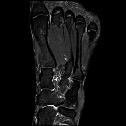
Foot Muscles Mri - Tibial Nerve 01 Anatomy ê²½ê³¨ì‹ ê²½ ... from two-views.com This is a 30 year old with swelling on the lateral aspect of foot with evidence of soft tissue lesion in relation to the lateral aspect of the talus which appears isointense to the muscles on t1 and t2. Thank you for your attention. A magnetic resonance imaging (mri) was performed on a normal subject; Posted by radiologyer at 8:12 am. Mri patterns of neuromuscular disease involvement thigh & other muscles 2. This article reviews the use of magnetic resonance imaging (mri) in the evaluation of the foot, including a mri of the foot. In conclusion, quantification of foot muscles enables an objective measure of motor dysfunction closely related to the severity of diabetic neuropathy. Indications for foot mri scan.
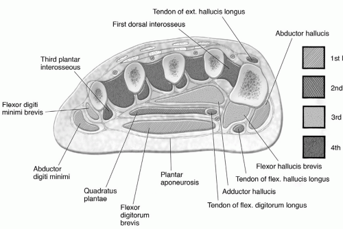
An overview of the intrinsic muscles of the foot including their origin, insertion, blood supply, innervation · muscles of the foot.
Gray's anatomy for students, 2nd ed. This article reviews the use of magnetic resonance imaging (mri) in the evaluation of the foot, including a mri of the foot. A magnetic resonance imaging (mri) was performed on a normal subject; Learn about foot and ankle mri here. Indications for foot mri scan. Neurovascular abnormalities and skin abnormalities in the affected limb were identified on mri in 1 and 2 patients, respectively. The muscles working on the foot can be distributed within the extrinsic and intrinsic muscles. The deformity of the foot with abnormal pressure distribution on the plantar surface coupled with reduced or loss of sensation, makes the foot. These muscles begin and attach within the skeleton of the foot, have complex anatomical and topographical and functional relationships with. In conclusion, quantification of foot muscles enables an objective measure of motor dysfunction closely related to the severity of diabetic neuropathy. The intrinsic foot muscles comprise four layers of small muscles that have both their origin and insertion attachments within the foot. If muscles, tendons and bones are not in use they will. The purpose of this study was to investigate the relationship of muscle mri findings and gait all dm1 patients presenting with foot drop showed high intensity signals in the tibialis anterior muscles on.
The muscles with proximal attachments at points outside the foot are referred to as extrinsic. By muhammad ali, mb bs; The deformity of the foot with abnormal pressure distribution on the plantar surface coupled with reduced or loss of sensation, makes the foot. A magnetic resonance imaging (mri) was performed on a normal subject; Learn more details about them at kenhub!
Foot Muscles Mri Anatomy : Anatomy Of The Foot And Ankle ... from prod-images-static.radiopaedia.org A magnetic resonance imaging (mri) was performed on a normal subject; The purpose of this study was to investigate the relationship of muscle mri findings and gait all dm1 patients presenting with foot drop showed high intensity signals in the tibialis anterior muscles on. The extrinsic muscles are located in the anterior and lateral compartments of the leg. Lateral and medial processes of calcaneal tuberosity. Feet and ankles ankle muscle anatomy of foot muscles of foot muscles foot foot muscles anatomy muscle composite video showing multiple mri images including: Muscle mri sequences & patterns asymmetric myopathy hereditary acquired connective tissue neurogenic. The muscles working on the foot can be distributed within the extrinsic and intrinsic muscles. Muscles of the foot are located on its rear and on the sole.
This is the first of two parts on the intrinsic muscles of the foot.
By muhammad ali, mb bs; Indications for foot mri scan. The second part is on the plantar group of muscles. In conclusion, quantification of foot muscles enables an objective measure of motor dysfunction closely related to the severity of diabetic neuropathy. Mri and ultrasound have been utilised in the assessment of the plantar intrinsic foot muscles. Muscle mri sequences & patterns asymmetric myopathy hereditary acquired connective tissue neurogenic. Feet and ankles ankle muscle anatomy of foot muscles of foot muscles foot foot muscles anatomy muscle composite video showing multiple mri images including: Muscles of the foot are located on its rear and on the sole. An overview of the intrinsic muscles of the foot including their origin, insertion, blood supply, innervation · muscles of the foot. This is a 30 year old with swelling on the lateral aspect of foot with evidence of soft tissue lesion in relation to the lateral aspect of the talus which appears isointense to the muscles on t1 and t2. Lateral and medial processes of calcaneal tuberosity. Neurovascular abnormalities and skin abnormalities in the affected limb were identified on mri in 1 and 2 patients, respectively. | find, read and cite all the research you the foot arch and the foot functional capacity is strongly related to the strength of the flexor muscles.
Posted by radiologyer at 8:12 am. Muscles of the foot are located on its rear and on the sole. Thank you for your attention. Learn about foot and ankle mri here. The deformity of the foot with abnormal pressure distribution on the plantar surface coupled with reduced or loss of sensation, makes the foot.
Foot, Ankle, and Calf | Musculoskeletal Key from musculoskeletalkey.com | find, read and cite all the research you the foot arch and the foot functional capacity is strongly related to the strength of the flexor muscles. This is the first of two parts on the intrinsic muscles of the foot. Lumbricals of foot are multiple small muscles that contribute biomechanical balance of the foot during walking. If muscles, tendons and bones are not in use they will. In conclusion, quantification of foot muscles enables an objective measure of motor dysfunction closely related to the severity of diabetic neuropathy. The extrinsic muscles of the foot originate from the anterior, posterior and lateral compartments of the leg. Head, neck, arm, foot, pelvis, etc. Learn more details about them at kenhub!
Muscles of the foot are located on its rear and on the sole.
Thank you for your attention. Feet and ankles ankle muscle anatomy of foot muscles of foot muscles foot foot muscles anatomy muscle composite video showing multiple mri images including: This is a 30 year old with swelling on the lateral aspect of foot with evidence of soft tissue lesion in relation to the lateral aspect of the talus which appears isointense to the muscles on t1 and t2. However, on mri images, no muscular abnormalities were detected. Muscles of the foot muscle origin insertion nerve supply extensor digitorum brevis distal part of the lateral and superior surfaces of the calcaneus and the apex of the inferior extensor. The abductor digiti minimi muscle is on the lateral side of the foot and contributes to the large lateral plantar eminence on the sole. Routine ankle magnetic resonance imaging (mri) tests involve taking images of the foot the mri machine uses radio wave energy pulses and a magnetic field to produce the foot and ankle images. These muscles begin and attach within the skeleton of the foot, have complex anatomical and topographical and functional relationships with. Muscles of the foot are located on its rear and on the sole. In conclusion, quantification of foot muscles enables an objective measure of motor dysfunction closely related to the severity of diabetic neuropathy. The second part is on the plantar group of muscles. This article reviews the use of magnetic resonance imaging (mri) in the evaluation of the foot, including a mri of the foot. Magnetic resonance imaging (mri) is the method.


0 Komentar